HLA-A*02:01 binding "RQFGPDWIVA" at 1.64Å resolution
Data provenance
Information sections
- Publication
- Peptide details
- Peptide neighbours
- Binding cleft pockets
- Chain sequences
- Downloadable data
- Data license
- Footnotes
Complex type
HLA-A*02:01
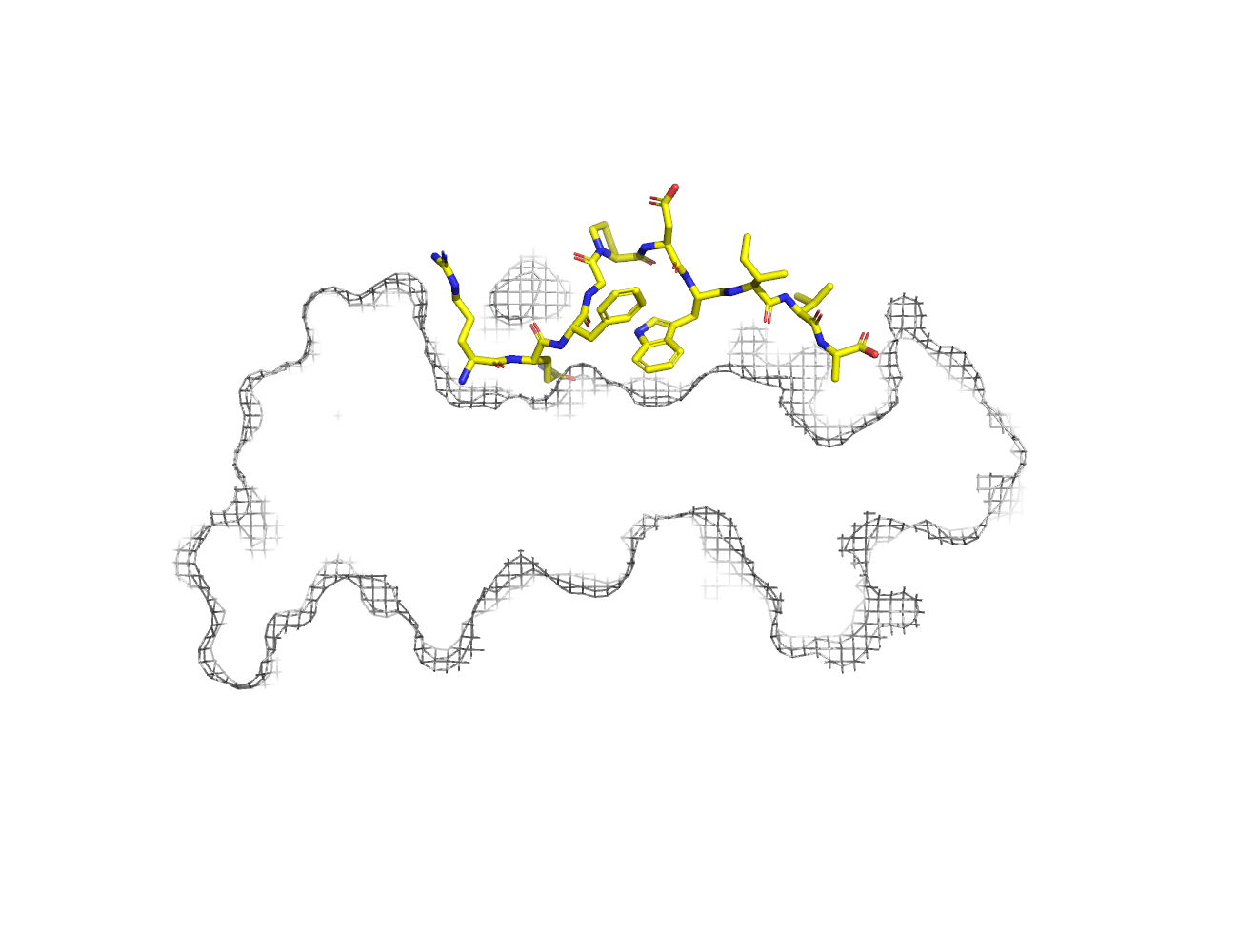
RQFGPDWIVA
Species
Locus / Allele group
Hotspot autoimmune T cell receptor binding underlies pathogen and insulin peptide cross-reactivity.
Structure deposition and release
Data provenance
Publication data retrieved from PDBe REST API8 and PMCe REST API9
Other structures from this publication


Data provenance
MHC:peptide complexes are visualised using PyMol. The peptide is superimposed on a consistent cutaway slice of the MHC binding cleft (displayed as a grey mesh) which best indicates the binding pockets for the P1/P5/PC positions (side view - pockets A, E, F) and for the P2/P3/PC-2 positions (top view - pockets B, C, D). In some cases peptides will use a different pocket for a specific peptide position (atypical anchoring). On some structures the peptide may appear to sterically clash with a pocket. This is an artefact of picking a standardised slice of the cleft and overlaying the peptide.
Peptide neighbours
|
P1
ARG
TYR159
THR163
GLU63
PHE33
MET5
TYR171
TYR7
TYR59
LYS66
TRP167
|
P10
ALA
THR142
ASP77
THR80
TRP147
THR143
TYR123
LYS146
TYR116
TYR84
|
P2
GLN
MET45
TYR99
PHE9
LYS66
VAL67
GLU63
TYR159
TYR7
HIS70
|
P3
PHE
LYS66
GLN155
TYR99
TYR159
LEU156
|
P4
GLY
LYS66
ARG65
|
P5
PRO
LYS66
ARG65
ALA69
|
P7
TRP
HIS114
TYR99
LYS66
ARG97
ALA69
THR73
LEU156
HIS70
|
P8
ILE
VAL152
GLN155
ARG97
ALA150
THR73
TRP147
|
P9
VAL
ASP77
THR73
LYS146
TRP147
VAL76
|
Colour key
Data provenance
Neighbours are calculated by finding residues with atoms within 5Å of each other using BioPython Neighboursearch module. The list of neighbours is then sorted and filtered to inlcude only neighbours where between the peptide and the MHC Class I alpha chain.
Colours selected to match the YRB scheme. [https://www.frontiersin.org/articles/10.3389/fmolb.2015.00056/full]


|
A Pocket
ALA159
GLY163
GLU167
ARG171
SER5
GLU59
GLY63
ARG66
ARG7
|
B Pocket
ILE24
PHE34
ARG45
GLY63
ARG66
LYS67
ARG7
ALA70
PHE9
MET99
|
C Pocket
ALA70
GLN73
THR74
PHE9
GLN97
|
D Pocket
TYR114
GLU155
GLN156
ALA159
TYR160
MET99
|
E Pocket
TYR114
LYS147
HIS152
GLN156
GLN97
|
F Pocket
GLN116
ASP123
THR143
HIS146
LYS147
VAL77
GLY80
THR81
GLY84
THR95
|
Colour key
Data provenance
|
1. Beta 2 microglobulin
Beta 2 microglobulin
|
10 20 30 40 50 60
MIQRTPKIQVYSRHPAENGKSNFLNCYVSGFHPSDIEVDLLKNGERIEKVEHSDLSFSKD 70 80 90 WSFYLLYYTEFTPTEKDEYACRVNHVTLSQPKIVKWDRDM |
|
2. Class I alpha
HLA-A*02:01
IPD-IMGT/HLA
[ipd-imgt:HLA35266] |
10 20 30 40 50 60
MGSHSMRYFFTSVSRPGRGEPRFIAVGYVDDTQFVRFDSDAASQRMEPRAPWIEQEGPEY 70 80 90 100 110 120 WDGETRKVKAHSQTHRVDLGTLRGYYNQSEAGSHTVQRMYGCDVGSDWRFLRGYHQYAYD 130 140 150 160 170 180 GKDYIALKEDLRSWTAADMAAQTTKHKWEAAHVAEQLRAYLEGTCVEWLRRYLENGKETL 190 200 210 220 230 240 QRTDAPKTHMTHHAVSDHEATLRCWALSFYPAEITLTWQRDGEDQTQDTELVETRPAGDG 250 260 270 TFQKWAAVVVPSGQEQRYTCHVQHEGLPKPLTLRWEP |
|
3. Peptide
|
RQFGPDWIVA
|
Data provenance
Sequences are retrieved via the Uniprot method of the RSCB REST API. Sequences are then compared to those derived from the PDB file and matched against sequences retrieved from the IPD-IMGT/HLA database for human sequences, or the IPD-MHC database for other species. Mouse sequences are matched against FASTA files from Uniprot. Sequences for the mature extracellular protein (signal petide and cytoplasmic tail removed) are compared to identical length sequences from the datasources mentioned before using either exact matching or Levenshtein distance based matching.
Downloadable data
Components
Data license
Footnotes
- Protein Data Bank Europe - Coordinate Server
- 1HHK - HLA-A*02:01 binding LLFGYPVYV at 2.5Å resolution - PDB entry for 1HHK
- Protein structure alignment by incremental combinatorial extension (CE) of the optimal path. - PyMol CEALIGN Method - Publication
- PyMol - PyMol.org/pymol
- Levenshtein distance - Wikipedia entry
- Protein Data Bank Europe REST API - Molecules endpoint
- 3Dmol.js: molecular visualization with WebGL - 3DMol.js - Publication
- Protein Data Bank Europe REST API - Publication endpoint
- PubMed Central Europe REST API - Articles endpoint

This work is licensed under a Creative Commons Attribution 4.0 International License.