H2-Db binding "YQLENYCGL" at 2.33Å resolution
Data provenance
Information sections
- Publication
- Peptide details
- Peptide neighbours
- Binding cleft pockets
- Chain sequences
- Downloadable data
- Data license
- Footnotes
Complex type
H2-Db
YQLENYCGL
Species
Locus / Allele group
Compensatory mechanisms allow undersized anchor-deficient class I MHC ligands to mediate pathogenic autoreactive T cell responses.
Self-reactive T cells must escape thymic negative selection to mediate pathogenic autoimmunity. In the NOD mouse model of autoimmune diabetes, several β cell-cytotoxic CD8 T cell populations are known, with the most aggressive of these represented by AI4, a T cell clone with promiscuous Ag-recognition characteristics. We identified a long-elusive β cell-specific ligand for AI4 as an unusually short H-2D(b)-binding 7-mer peptide lacking a C-terminal anchor residue and derived from the insulin A chain (InsA14-20). Crystallography reveals that compensatory mechanisms permit peptides lacking a C-terminal anchor to bind sufficiently to the MHC to enable destructive T cell responses, yet allow cognate T cells to avoid negative selection. InsA14-20 shares two solvent-exposed residues with previously identified AI4 ligands, providing a structural explanation for AI4's promiscuity. Detection of AI4-like T cells, using mimotopes of InsA14-20 with improved H-2D(b)-binding characteristics, establishes the AI4-like T cell population as a consistent feature of the islet infiltrates of NOD mice. Our work establishes undersized peptides as previously unrecognized targets of autoreactive CD8 T cells and presents a strategy for their further exploration as Ags in autoimmune disease.
Structure deposition and release
Data provenance
Publication data retrieved from PDBe REST API8 and PMCe REST API9
Other structures from this publication


Data provenance
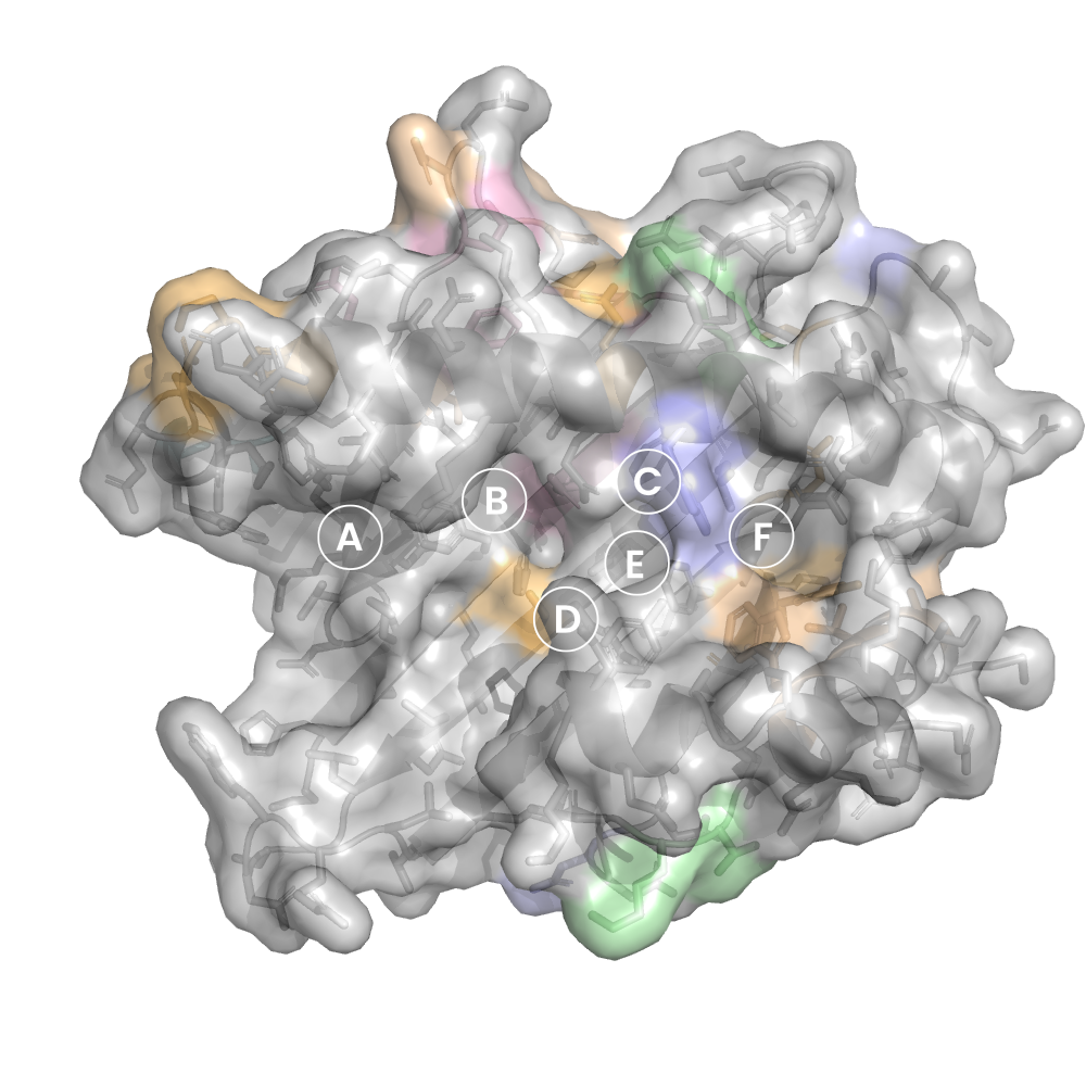
MHC:peptide complexes are visualised using PyMol. The peptide is superimposed on a consistent cutaway slice of the MHC binding cleft (displayed as a grey mesh) which best indicates the binding pockets for the P1/P5/PC positions (side view - pockets A, E, F) and for the P2/P3/PC-2 positions (top view - pockets B, C, D). In some cases peptides will use a different pocket for a specific peptide position (atypical anchoring). On some structures the peptide may appear to sterically clash with a pocket. This is an artefact of picking a standardised slice of the cleft and overlaying the peptide.
Peptide neighbours
|
P1
TYR
TYR183
TYR31
TYR195
PHE57
ARG86
TYR83
GLU187
MET29
LYS90
TRP191
GLU87
|
P2
GLN
GLU33
SER48
TYR31
LYS90
GLU87
ALA91
TYR183
TYR46
TYR69
GLN94
|
P3
LEU
LEU138
GLN94
GLN121
HIS179
SER123
LYS90
TYR183
TYR180
|
P4
GLU
HIS179
LYS90
TYR180
GLN94
|
P5
ASN
PHE140
HIS179
GLN94
GLU33
GLN121
TYR180
PHE98
TRP97
|
P6
TYR
TYR180
TRP97
GLY175
SER174
ALA176
HIS179
|
P7
CYS
TYR180
TRP171
TRP97
SER174
ALA176
|
P8
GLY
TRP171
TRP97
THR167
SER101
LYS170
|
P9
LEU
LYS170
TRP171
TYR108
TRP97
LEU119
SER101
THR167
LEU105
ASN104
PHE140
TYR147
ILE148
|
Colour key
Data provenance
Neighbours are calculated by finding residues with atoms within 5Å of each other using BioPython Neighboursearch module. The list of neighbours is then sorted and filtered to inlcude only neighbours where between the peptide and the MHC Class I alpha chain.
Colours selected to match the YRB scheme. [https://www.frontiersin.org/articles/10.3389/fmolb.2015.00056/full]

|
A Pocket
LEU159
CYS163
LEU167
LEU171
ARG5
TRP59
THR63
ALA66
PHE7
|
B Pocket
VAL24
ARG34
GLU45
THR63
ALA66
LYS67
PHE7
GLU70
THR9
GLY99
|
C Pocket
GLU70
PHE73
ARG74
THR9
MET97
|
D Pocket
GLN114
TYR155
LYS156
LEU159
GLU160
GLY99
|
E Pocket
GLN114
GLU147
ALA152
LYS156
MET97
|
F Pocket
ALA116
ILE123
ARG143
TRP146
GLU147
LEU77
LEU80
LEU81
TYR84
GLN95
|
Colour key
Data provenance
|
1. Beta 2 microglobulin
Beta 2 microglobulin
|
10 20 30 40 50 60
MIQKTPQIQVYSRHPPENGKPNILNCYVTQFHPPHIEIQMLKNGKKIPKVEMSDMSFSKD 70 80 90 WSFYILAHTEFTPTETDTYACRVKHDSMAEPKTVYWDRDM |
|
2. Class I alpha
H2-Db
|
10 20 30 40 50 60
PHSMRYFETAVSRPGLEEPRYISVGYVDNKEFVRFDSDAENPRYEPRAPWMEQEGPEYWE 70 80 90 100 110 120 RETQKAKGQEQWFRVSLRNLLGYYNQSAGGSHTLQQMSGCDLGSDWRLLRGYLQFAYEGR 130 140 150 160 170 180 DYIALNEDLKTWTAADMAAQITRRKWEQSGAAEHYKAYLEGECVEWLHRYLKNGNATLLR 190 200 210 220 230 240 TDSPKAHVTHHPRSKGEVTLRCWALGFYPADITLTWQLNGEELTQDMELVETRPAGDGTF 250 260 270 QKWASVVVPLGKEQNYTCRVYHEGLPEPLTLRW |
|
3. Peptide
|
YQLENYCGL
|
Data provenance
Sequences are retrieved via the Uniprot method of the RSCB REST API. Sequences are then compared to those derived from the PDB file and matched against sequences retrieved from the IPD-IMGT/HLA database for human sequences, or the IPD-MHC database for other species. Mouse sequences are matched against FASTA files from Uniprot. Sequences for the mature extracellular protein (signal petide and cytoplasmic tail removed) are compared to identical length sequences from the datasources mentioned before using either exact matching or Levenshtein distance based matching.
Downloadable data
Components
Data license
Footnotes
- Protein Data Bank Europe - Coordinate Server
- 1HHK - HLA-A*02:01 binding LLFGYPVYV at 2.5Å resolution - PDB entry for 1HHK
- Protein structure alignment by incremental combinatorial extension (CE) of the optimal path. - PyMol CEALIGN Method - Publication
- PyMol - PyMol.org/pymol
- Levenshtein distance - Wikipedia entry
- Protein Data Bank Europe REST API - Molecules endpoint
- 3Dmol.js: molecular visualization with WebGL - 3DMol.js - Publication
- Protein Data Bank Europe REST API - Publication endpoint
- PubMed Central Europe REST API - Articles endpoint

This work is licensed under a Creative Commons Attribution 4.0 International License.