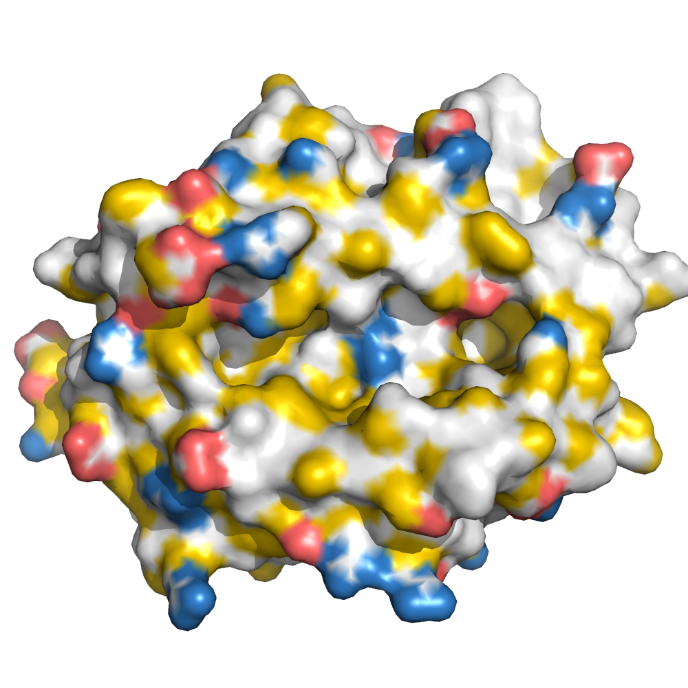
HLA-A*02:01 binding "YMFPNAPYL" at 1.89Å resolution
Data provenance
Information sections
- Publication
- Peptide details
- Peptide neighbours
- Binding cleft pockets
- Chain sequences
- Downloadable data
- Data license
- Footnotes
Complex type
HLA-A*02:01
YMFPNAPYL
Species
Locus / Allele group
Structures of native and affinity-enhanced WT1 epitopes bound to HLA-A*0201: implications for WT1-based cancer therapeutics.
Presentation of peptides by class I or class II major histocompatibility complex (MHC) molecules is required for the initiation and propagation of a T cell-mediated immune response. Peptides from the Wilms Tumor 1 transcription factor (WT1), upregulated in many hematopoetic and solid tumors, can be recognized by T cells and numerous efforts are underway to engineer WT1-based cancer vaccines. Here we determined the structures of the class I MHC molecule HLA-A*0201 bound to the native 126-134 epitope of the WT1 peptide and a recently described variant (R1Y) with improved MHC binding. The R1Y variant, a potential vaccine candidate, alters the positions of MHC charged side chains near the peptide N-terminus and significantly reduces the peptide/MHC electrostatic surface potential. These alterations indicate that the R1Y variant is an imperfect mimic of the native WT1 peptide, and suggest caution in its use as a therapeutic vaccine. Stability measurements revealed how the R1Y substitution enhances MHC binding affinity, and together with the structures suggest a strategy for engineering WT1 variants with improved MHC binding that retain the structural features of the native peptide/MHC complex.
Structure deposition and release
Data provenance
Publication data retrieved from PDBe REST API8 and PMCe REST API9
Other structures from this publication


Data provenance
MHC:peptide complexes are visualised using PyMol. The peptide is superimposed on a consistent cutaway slice of the MHC binding cleft (displayed as a grey mesh) which best indicates the binding pockets for the P1/P5/PC positions (side view - pockets A, E, F) and for the P2/P3/PC-2 positions (top view - pockets B, C, D). In some cases peptides will use a different pocket for a specific peptide position (atypical anchoring). On some structures the peptide may appear to sterically clash with a pocket. This is an artefact of picking a standardised slice of the cleft and overlaying the peptide.
Peptide neighbours
|
P1
TYR
TRP167
TYR159
TYR59
PHE33
MET5
TYR171
THR163
TYR7
LYS66
GLU63
|
P2
MET
GLU63
MET45
TYR7
TYR99
HIS70
LYS66
VAL67
TYR159
PHE9
|
P3
PHE
TYR159
HIS70
LYS66
TYR99
LEU156
GLN155
ARG97
|
P4
PRO
HIS70
LYS66
TYR159
|
P5
ASN
HIS70
GLN155
ARG97
|
P6
ALA
TYR116
THR73
ARG97
HIS70
HIS74
|
P7
PRO
VAL152
ASP77
TYR116
THR73
ARG97
TRP147
HIS114
|
P8
TYR
THR143
LYS146
VAL76
THR73
ASP77
TRP147
|
P9
LEU
THR143
LYS146
TYR84
TYR116
TYR123
VAL95
THR80
TRP147
LEU81
THR142
ASP77
ILE124
|
Colour key
Data provenance
Neighbours are calculated by finding residues with atoms within 5Å of each other using BioPython Neighboursearch module. The list of neighbours is then sorted and filtered to inlcude only neighbours where between the peptide and the MHC Class I alpha chain.
Colours selected to match the YRB scheme. [https://www.frontiersin.org/articles/10.3389/fmolb.2015.00056/full]


|
A Pocket
TYR159
THR163
TRP167
TYR171
MET5
TYR59
GLU63
LYS66
TYR7
|
B Pocket
ALA24
VAL34
MET45
GLU63
LYS66
VAL67
TYR7
HIS70
PHE9
TYR99
|
C Pocket
HIS70
THR73
HIS74
PHE9
ARG97
|
D Pocket
HIS114
GLN155
LEU156
TYR159
LEU160
TYR99
|
E Pocket
HIS114
TRP147
VAL152
LEU156
ARG97
|
F Pocket
TYR116
TYR123
THR143
LYS146
TRP147
ASP77
THR80
LEU81
TYR84
VAL95
|
Colour key
Data provenance
|
1. Beta 2 microglobulin
Beta 2 microglobulin
|
10 20 30 40 50 60
MIQRTPKIQVYSRHPAENGKSNFLNCYVSGFHPSDIEVDLLKNGERIEKVEHSDLSFSKD 70 80 90 WSFYLLYYTEFTPTEKDEYACRVNHVTLSQPKIVKWDRDM |
|
2. Class I alpha
HLA-A*02:01
IPD-IMGT/HLA
[ipd-imgt:HLA35266] |
10 20 30 40 50 60
GSHSMRYFFTSVSRPGRGEPRFIAVGYVDDTQFVRFDSDAASQRMEPRAPWIEQEGPEYW 70 80 90 100 110 120 DGETRKVKAHSQTHRVDLGTLRGYYNQSEAGSHTVQRMYGCDVGSDWRFLRGYHQYAYDG 130 140 150 160 170 180 KDYIALKEDLRSWTAADMAAQTTKHKWEAAHVAEQLRAYLEGTCVEWLRRYLENGKETLQ 190 200 210 220 230 240 RTDAPKTHMTHHAVSDHEATLRCWALSFYPAEITLTWQRDGEDQTQDTELVETRPAGDGT 250 260 270 FQKWAAVVVPSGQEQRYTCHVQHEGLPKPLTLRWE |
|
3. Peptide
|
YMFPNAPYL
|
Data provenance
Sequences are retrieved via the Uniprot method of the RSCB REST API. Sequences are then compared to those derived from the PDB file and matched against sequences retrieved from the IPD-IMGT/HLA database for human sequences, or the IPD-MHC database for other species. Mouse sequences are matched against FASTA files from Uniprot. Sequences for the mature extracellular protein (signal petide and cytoplasmic tail removed) are compared to identical length sequences from the datasources mentioned before using either exact matching or Levenshtein distance based matching.
Downloadable data
Components
Data license
Footnotes
- Protein Data Bank Europe - Coordinate Server
- 1HHK - HLA-A*02:01 binding LLFGYPVYV at 2.5Å resolution - PDB entry for 1HHK
- Protein structure alignment by incremental combinatorial extension (CE) of the optimal path. - PyMol CEALIGN Method - Publication
- PyMol - PyMol.org/pymol
- Levenshtein distance - Wikipedia entry
- Protein Data Bank Europe REST API - Molecules endpoint
- 3Dmol.js: molecular visualization with WebGL - 3DMol.js - Publication
- Protein Data Bank Europe REST API - Publication endpoint
- PubMed Central Europe REST API - Articles endpoint

This work is licensed under a Creative Commons Attribution 4.0 International License.