H2-Kb binding "RAQIFANI" at 1.80Å resolution
Data provenance
Information sections
- Publication
- Peptide details
- Peptide neighbours
- Binding cleft pockets
- Chain sequences
- Downloadable data
- Data license
- Footnotes
Complex type
H2-Kb
RAQIFANI
Species
Locus / Allele group
Prevention of cytotoxic T cell escape using a heteroclitic subdominant viral T cell determinant.
High affinity antigen-specific T cells play a critical role during protective immune responses. Epitope enhancement can elicit more potent T cell responses and can subsequently lead to a stronger memory pool; however, the molecular basis of such enhancement is unclear. We used the consensus peptide-binding motif for the Major Histocompatibility Complex molecule H-2K(b) to design a heteroclitic version of the mouse hepatitis virus-specific subdominant S598 determinant. We demonstrate that a single amino acid substitution at a secondary anchor residue (Q to Y at position 3) increased the stability of the engineered determinant in complex with H-2K(b). The structural basis for this enhanced stability was associated with local alterations in the pMHC conformation as a result of the Q to Y substitution. Recombinant viruses encoding this engineered determinant primed CTL responses that also reacted to the wildtype epitope with significantly higher functional avidity, and protected against selection of virus mutated at a second CTL determinant and consequent disease progression in persistently infected mice. Collectively, our findings provide a basis for the enhanced immunogenicity of an engineered determinant that will serve as a template for guiding the development of heteroclitic T cell determinants with applications in prevention of CTL escape in chronic viral infections as well as in tumor immunity.
Structure deposition and release
Data provenance
Publication data retrieved from PDBe REST API8 and PMCe REST API9
Other structures from this publication


Data provenance
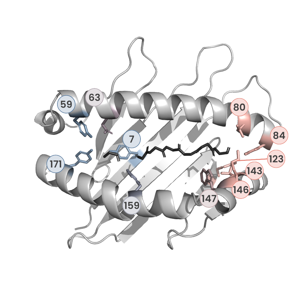
MHC:peptide complexes are visualised using PyMol. The peptide is superimposed on a consistent cutaway slice of the MHC binding cleft (displayed as a grey mesh) which best indicates the binding pockets for the P1/P5/PC positions (side view - pockets A, E, F) and for the P2/P3/PC-2 positions (top view - pockets B, C, D). In some cases peptides will use a different pocket for a specific peptide position (atypical anchoring). On some structures the peptide may appear to sterically clash with a pocket. This is an artefact of picking a standardised slice of the cleft and overlaying the peptide.
Peptide neighbours
|
P1
ARG
TRP167
LEU5
GLU63
TYR171
TYR159
TYR59
LYS66
TYR7
THR163
|
P3
GLN
GLN114
GLU152
ASN70
TYR159
LYS66
LEU156
SER99
ARG155
|
P4
ILE
ASN70
ARG155
LYS66
|
P5
PHE
ASN70
VAL9
ARG155
GLN114
SER99
GLU24
TYR116
VAL97
PHE74
TYR22
SER73
|
P6
ALA
ASP77
TYR116
GLU152
ARG155
TRP147
|
P7
ASN
ASP77
VAL76
LYS146
SER73
TRP147
|
P8
ILE
THR80
ASP77
THR143
TYR116
LYS146
TYR123
LEU81
TRP147
TYR84
|
Colour key
Data provenance
Neighbours are calculated by finding residues with atoms within 5Å of each other using BioPython Neighboursearch module. The list of neighbours is then sorted and filtered to inlcude only neighbours where between the peptide and the MHC Class I alpha chain.
Colours selected to match the YRB scheme. [https://www.frontiersin.org/articles/10.3389/fmolb.2015.00056/full]

|
A Pocket
TYR159
THR163
TRP167
TYR171
LEU5
TYR59
GLU63
LYS66
TYR7
|
B Pocket
GLU24
VAL34
TYR45
GLU63
LYS66
ALA67
TYR7
ASN70
VAL9
SER99
|
C Pocket
ASN70
SER73
PHE74
VAL9
VAL97
|
D Pocket
GLN114
ARG155
LEU156
TYR159
LEU160
SER99
|
E Pocket
GLN114
TRP147
GLU152
LEU156
VAL97
|
F Pocket
TYR116
TYR123
THR143
LYS146
TRP147
ASP77
THR80
LEU81
TYR84
ILE95
|
Colour key
Data provenance
|
1. Beta 2 microglobulin
Beta 2 microglobulin
|
10 20 30 40 50 60
IQKTPQIQVYSRHPPENGKPNILNCYVTQFHPPHIEIQMLKNGKKIPKVEMSDMSFSKDW 70 80 90 SFYILAHTEFTPTETDTYACRVKHDSMAEPKTVYWDRDM |
|
2. Class I alpha
H2-Kb
|
10 20 30 40 50 60
GPHSLRYFVTAVSRPGLGEPRYMEVGYVDDTEFVRFDSDAENPRYEPRARWMEQEGPEYW 70 80 90 100 110 120 ERETQKAKGNEQSFRVDLRTLLGYYNQSKGGSHTIQVISGCEVGSDGRLLRGYQQYAYDG 130 140 150 160 170 180 CDYIALNEDLKTWTAADMAALITKHKWEQAGEAERLRAYLEGTCVEWLRRYLKNGNATLL 190 200 210 220 230 240 RTDSPKAHVTHHSRPEDKVTLRCWALGFYPADITLTWQLNGEELIQDMELVETRPAGDGT 250 260 270 FQKWASVVVPLGKEQYYTCHVYHQGLPEPLTLRWEPPP |
|
3. Peptide
|
RAQIFANI
|
Data provenance
Sequences are retrieved via the Uniprot method of the RSCB REST API. Sequences are then compared to those derived from the PDB file and matched against sequences retrieved from the IPD-IMGT/HLA database for human sequences, or the IPD-MHC database for other species. Mouse sequences are matched against FASTA files from Uniprot. Sequences for the mature extracellular protein (signal petide and cytoplasmic tail removed) are compared to identical length sequences from the datasources mentioned before using either exact matching or Levenshtein distance based matching.
Downloadable data
Components
Data license
Footnotes
- Protein Data Bank Europe - Coordinate Server
- 1HHK - HLA-A*02:01 binding LLFGYPVYV at 2.5Å resolution - PDB entry for 1HHK
- Protein structure alignment by incremental combinatorial extension (CE) of the optimal path. - PyMol CEALIGN Method - Publication
- PyMol - PyMol.org/pymol
- Levenshtein distance - Wikipedia entry
- Protein Data Bank Europe REST API - Molecules endpoint
- 3Dmol.js: molecular visualization with WebGL - 3DMol.js - Publication
- Protein Data Bank Europe REST API - Publication endpoint
- PubMed Central Europe REST API - Articles endpoint

This work is licensed under a Creative Commons Attribution 4.0 International License.