H2-Db binding "ASLWNGPHL" at 2.10Å resolution
Data provenance
Information sections
- Publication
- Peptide details
- Peptide neighbours
- Binding cleft pockets
- Chain sequences
- Downloadable data
- Data license
- Footnotes
Complex type
H2-Db
ASLWNGPHL
Species
Locus / Allele group
Structural and biological basis of CTL escape in coronavirus-infected mice.
Cytotoxic T lymphocyte escape occurs in many human infections, as well as mice infected with the JHM strain of mouse hepatitis virus, which exhibit CTL escape variants with mutations in a single epitope from the spike glycoprotein (S510). In all CTL epitopes prone to escape, only a subset of all potential variants is generally detected, even though many of the changes that are not selected would result in evasion of the T cell response. It is postulated that these unselected mutations significantly impair virus fitness. To define more precisely the basis for this preferential selection, we combine x-ray crystallographic studies of the MHC class I (D(b))/S510 complexes with viral reverse genetics to identify a prominent TCR contact residue (tryptophan at position 4) prone to escape mutations. The data show that a mutation that is commonly detected in chronically infected mice (tryptophan to arginine) potently disrupts the topology of the complex, explaining its selection. However, other mutations at this residue, which also abrogate the CTL response, are never selected in vivo even though they do not compromise virus fitness in acutely infected animals or induce a significant de novo CTL response. Thus, while structural analyses of the S510/D(b) complex provide a strong basis for why some CTL escape variants are selected, our results also show that factors other than effects on virus fitness limit the diversification of CD8 T cell epitopes.
Structure deposition and release
Data provenance
Publication data retrieved from PDBe REST API8 and PMCe REST API9
Other structures from this publication


Data provenance
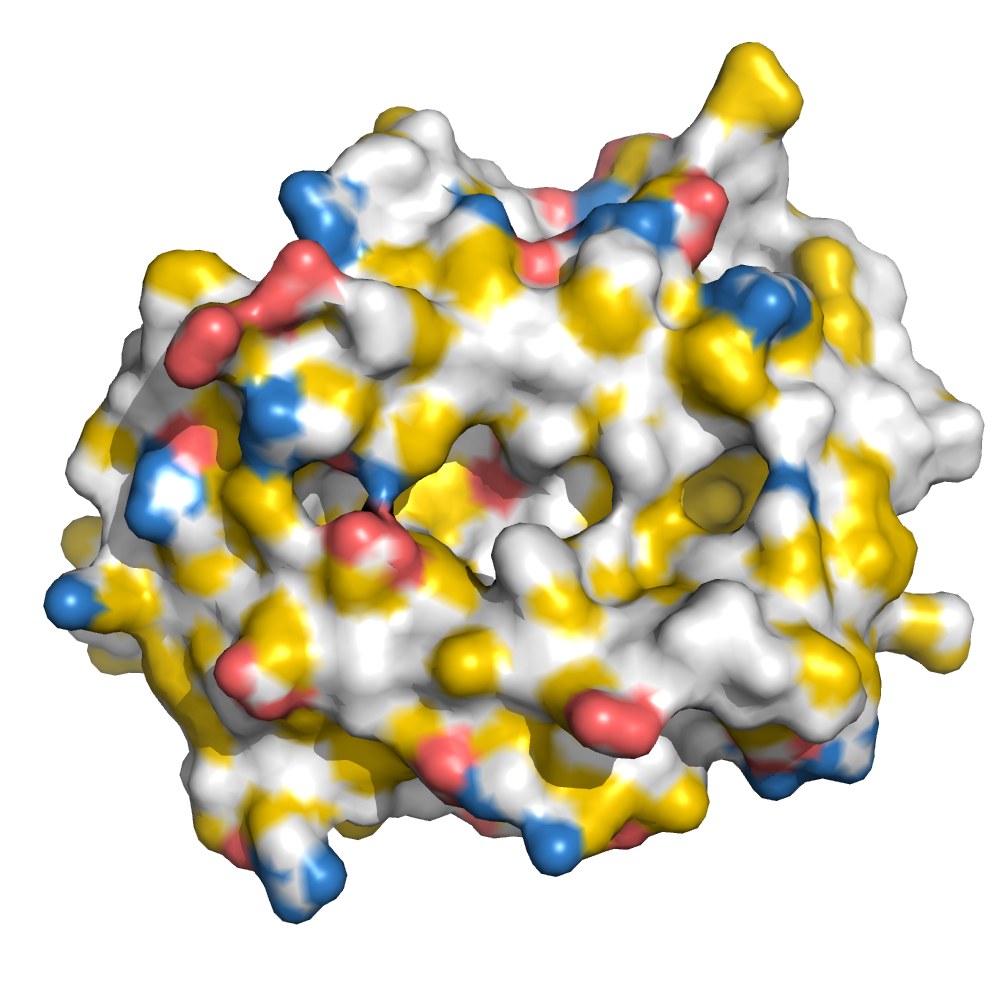
MHC:peptide complexes are visualised using PyMol. The peptide is superimposed on a consistent cutaway slice of the MHC binding cleft (displayed as a grey mesh) which best indicates the binding pockets for the P1/P5/PC positions (side view - pockets A, E, F) and for the P2/P3/PC-2 positions (top view - pockets B, C, D). In some cases peptides will use a different pocket for a specific peptide position (atypical anchoring). On some structures the peptide may appear to sterically clash with a pocket. This is an artefact of picking a standardised slice of the cleft and overlaying the peptide.
Peptide neighbours
|
P2
SER
TYR45
TYR159
GLU163
GLU63
LYS66
TYR7
|
P3
LEU
TYR156
GLN70
TYR159
SER99
LYS66
LEU114
GLU9
GLN97
|
P4
TRP
GLN70
GLY69
LYS66
HIS155
GLN65
TYR156
|
P5
ASN
PHE116
TRP73
LEU114
GLN70
HIS155
PHE74
GLN97
TYR156
|
P6
GLY
TYR156
TRP73
HIS155
|
P7
PRO
ALA152
SER150
TRP147
LYS146
TYR156
TRP73
|
P8
HIS
TRP147
GLN72
LYS146
THR143
SER77
TRP73
VAL76
ASN80
|
P9
LEU
SER77
TRP147
PHE116
TRP73
TYR84
TYR123
LEU95
LYS146
ILE124
THR143
LEU81
ASN80
|
Colour key
Data provenance
Neighbours are calculated by finding residues with atoms within 5Å of each other using BioPython Neighboursearch module. The list of neighbours is then sorted and filtered to inlcude only neighbours where between the peptide and the MHC Class I alpha chain.
Colours selected to match the YRB scheme. [https://www.frontiersin.org/articles/10.3389/fmolb.2015.00056/full]


|
A Pocket
TYR159
GLU163
TRP167
TYR171
MET5
TYR59
GLU63
LYS66
TYR7
|
B Pocket
SER24
VAL34
TYR45
GLU63
LYS66
ALA67
TYR7
GLN70
GLU9
SER99
|
C Pocket
GLN70
TRP73
PHE74
GLU9
GLN97
|
D Pocket
LEU114
HIS155
TYR156
TYR159
LEU160
SER99
|
E Pocket
LEU114
TRP147
ALA152
TYR156
GLN97
|
F Pocket
PHE116
TYR123
THR143
LYS146
TRP147
SER77
ASN80
LEU81
TYR84
LEU95
|
Colour key
Data provenance
|
1. Beta 2 microglobulin
Beta 2 microglobulin
|
10 20 30 40 50 60
MIQKTPQIQVYSRHPPENGKPNILNCYVTQFHPPHIEIQMLKNGKKIPKVEMSDMSFSKD 70 80 90 WSFYILAHTEFTPTETDTYACRVKHDSMAEPKTVYWDRDM |
|
2. Class I alpha
H2-Db
|
10 20 30 40 50 60
GPHSMRYFETAVSRPGLEEPRYISVGYVDNKEFVRFDSDAENPRYEPRAPWMEQEGPEYW 70 80 90 100 110 120 ERETQKAKGQEQWFRVSLRNLLGYYNQSAGGSHTLQQMSGCDLGSDWRLLRGYLQFAYEG 130 140 150 160 170 180 RDYIALNEDLKTWTAADMAAQITRRKWEQSGAAEHYKAYLEGECVEWLHRYLKNGNATLL 190 200 210 220 230 240 RTDSPKAHVTHHPRSKGEVTLRCWALGFYPADITLTWQLNGEELTQDMELVETRPAGDGT 250 260 270 FQKWASVVVPLGKEQNYTCRVYHEGLPEPLTLRWERWE |
|
3. Peptide
|
ASLWNGPHL
|
Data provenance
Sequences are retrieved via the Uniprot method of the RSCB REST API. Sequences are then compared to those derived from the PDB file and matched against sequences retrieved from the IPD-IMGT/HLA database for human sequences, or the IPD-MHC database for other species. Mouse sequences are matched against FASTA files from Uniprot. Sequences for the mature extracellular protein (signal petide and cytoplasmic tail removed) are compared to identical length sequences from the datasources mentioned before using either exact matching or Levenshtein distance based matching.
Downloadable data
Complete structures
Components
Data license
Footnotes
- Protein Data Bank Europe - Coordinate Server
- 1HHK - HLA-A*02:01 binding LLFGYPVYV at 2.5Å resolution - PDB entry for 1HHK
- Protein structure alignment by incremental combinatorial extension (CE) of the optimal path. - PyMol CEALIGN Method - Publication
- PyMol - PyMol.org/pymol
- Levenshtein distance - Wikipedia entry
- Protein Data Bank Europe REST API - Molecules endpoint
- 3Dmol.js: molecular visualization with WebGL - 3DMol.js - Publication
- Protein Data Bank Europe REST API - Publication endpoint
- PubMed Central Europe REST API - Articles endpoint

This work is licensed under a Creative Commons Attribution 4.0 International License.