HLA-A*02:01 binding "MILGGVFXV" at 2.30Å resolution
Data provenance
Information sections
- Publication
- Peptide details
- Peptide neighbours
- Binding cleft pockets
- Chain sequences
- Downloadable data
- Data license
- Footnotes
Complex type
HLA-A*02:01
MILGGVFXV
Species
Locus / Allele group
Class I major histocompatibility complexes loaded by a periodate trigger.
Class I major histocompatibility complexes (MHCs) present peptide ligands on the cell surface for recognition by appropriate cytotoxic T cells. The unstable nature of unliganded MHC necessitates the production of recombinant class I complexes through in vitro refolding reactions in the presence of an added excess of peptides. This strategy is not amenable to high-throughput production of vast collections of class I complexes. To address this issue, we recently designed photocaged MHC ligands that can be cleaved by a UV light trigger in the MHC bound state under conditions that do not affect the integrity of the MHC structure. The results obtained with photocaged MHC ligands demonstrate that conditional MHC ligands can form a generally applicable concept for the creation of defined peptide-MHCs. However, the use of UV exposure to mediate ligand exchange is unsuited for a number of applications, due to the lack of UV penetration through cell culture systems and due to the transfer of heat upon UV irradiation, which can induce evaporation. To overcome these limitations, here, we provide proof-of-concept for the generation of defined peptide-MHCs by chemical trigger-induced ligand exchange. The crystal structure of the MHC with the novel chemosensitive ligand showcases that the ligand occupies the expected binding site, in a conformation where the hydroxyl groups should be reactive to periodate. We proceed to validate this technology by producing peptide-MHCs that can be used for T cell detection. The methodology that we describe here should allow loading of MHCs with defined peptides in cell culture devices, thereby permitting antigen-specific T cell expansion and purification for cell therapy. In addition, this technology will be useful to develop miniaturized assay systems for performing high-throughput screens for natural and unnatural MHC ligands.
Structure deposition and release
Data provenance
Publication data retrieved from PDBe REST API8 and PMCe REST API9
Other structures from this publication


Data provenance
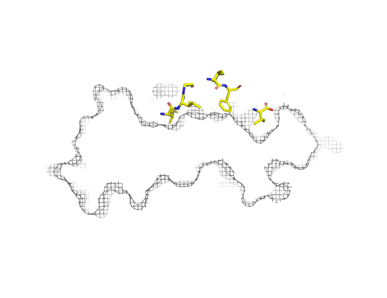
MHC:peptide complexes are visualised using PyMol. The peptide is superimposed on a consistent cutaway slice of the MHC binding cleft (displayed as a grey mesh) which best indicates the binding pockets for the P1/P5/PC positions (side view - pockets A, E, F) and for the P2/P3/PC-2 positions (top view - pockets B, C, D). In some cases peptides will use a different pocket for a specific peptide position (atypical anchoring). On some structures the peptide may appear to sterically clash with a pocket. This is an artefact of picking a standardised slice of the cleft and overlaying the peptide.
Peptide neighbours
|
P2
ILE
GLU63
VAL67
TYR159
TYR7
PHE9
HIS70
TYR99
THR163
MET45
LYS66
|
P3
LEU
GLN155
TYR159
LEU156
ARG97
HIS70
TYR99
LYS66
|
P4
GLY
HIS70
ALA69
LYS66
|
P6
VAL
GLN155
|
P7
PHE
TRP147
ARG97
THR73
HIS114
VAL152
LEU156
|
P9
VAL
TYR123
THR80
TYR116
TRP147
LEU81
ASP77
TYR84
LYS146
THR143
|
Colour key
Data provenance
Neighbours are calculated by finding residues with atoms within 5Å of each other using BioPython Neighboursearch module. The list of neighbours is then sorted and filtered to inlcude only neighbours where between the peptide and the MHC Class I alpha chain.
Colours selected to match the YRB scheme. [https://www.frontiersin.org/articles/10.3389/fmolb.2015.00056/full]


|
A Pocket
TYR159
THR163
TRP167
TYR171
MET5
TYR59
GLU63
LYS66
TYR7
|
B Pocket
ALA24
VAL34
MET45
GLU63
LYS66
VAL67
TYR7
HIS70
PHE9
TYR99
|
C Pocket
HIS70
THR73
HIS74
PHE9
ARG97
|
D Pocket
HIS114
GLN155
LEU156
TYR159
LEU160
TYR99
|
E Pocket
HIS114
TRP147
VAL152
LEU156
ARG97
|
F Pocket
TYR116
TYR123
THR143
LYS146
TRP147
ASP77
THR80
LEU81
TYR84
VAL95
|
Colour key
Data provenance
|
1. Beta 2 microglobulin
Beta 2 microglobulin
|
10 20 30 40 50 60
MIQRTPKIQVYSRHPAENGKSNFLNCYVSGFHPSDIEVDLLKNGERIEKVEHSDLSFSKD 70 80 90 WSFYLLYYTEFTPTEKDEYACRVNHVTLSQPKIVKWDRDM |
|
2. Class I alpha
HLA-A*02:01
IPD-IMGT/HLA
[ipd-imgt:HLA35266] |
10 20 30 40 50 60
GSHSMRYFFTSVSRPGRGEPRFIAVGYVDDTQFVRFDSDAASQRMEPRAPWIEQEGPEYW 70 80 90 100 110 120 DGETRKVKAHSQTHRVDLGTLRGYYNQSEAGSHTVQRMYGCDVGSDWRFLRGYHQYAYDG 130 140 150 160 170 180 KDYIALKEDLRSWTAADMAAQTTKHKWEAAHVAEQLRAYLEGTCVEWLRRYLENGKETLQ 190 200 210 220 230 240 RTDAPKTHMTHHAVSDHEATLRCWALSFYPAEITLTWQRDGEDQTQDTELVETRPAGDGT 250 260 270 FQKWAAVVVPSGQEQRYTCHVQHEGLPKPLTLRWE |
|
3. Peptide
|
MILGGVFXV
|
Data provenance
Sequences are retrieved via the Uniprot method of the RSCB REST API. Sequences are then compared to those derived from the PDB file and matched against sequences retrieved from the IPD-IMGT/HLA database for human sequences, or the IPD-MHC database for other species. Mouse sequences are matched against FASTA files from Uniprot. Sequences for the mature extracellular protein (signal petide and cytoplasmic tail removed) are compared to identical length sequences from the datasources mentioned before using either exact matching or Levenshtein distance based matching.
Downloadable data
Components
Data license
Footnotes
- Protein Data Bank Europe - Coordinate Server
- 1HHK - HLA-A*02:01 binding LLFGYPVYV at 2.5Å resolution - PDB entry for 1HHK
- Protein structure alignment by incremental combinatorial extension (CE) of the optimal path. - PyMol CEALIGN Method - Publication
- PyMol - PyMol.org/pymol
- Levenshtein distance - Wikipedia entry
- Protein Data Bank Europe REST API - Molecules endpoint
- 3Dmol.js: molecular visualization with WebGL - 3DMol.js - Publication
- Protein Data Bank Europe REST API - Publication endpoint
- PubMed Central Europe REST API - Articles endpoint

This work is licensed under a Creative Commons Attribution 4.0 International License.